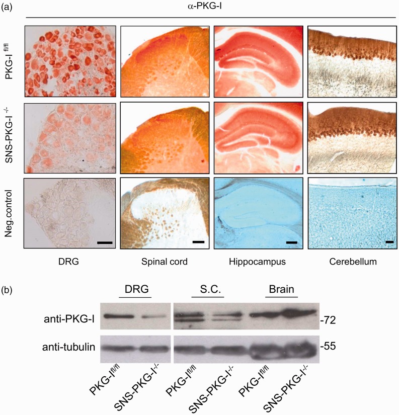
Figure 2.
Identification of specific deletion of PKG-I in nociceptors in SNS-PKG- I−/− mice. (a) Immunohistochemical staining showing that PKG-I is specifically deleted in small- to medium-diameter DRG neurons and superficial lamina of spinal dorsal horn where nociceptive primary afferents mainly terminates, but without alterations in the brain regions, such as hippocampus and cerebellum. (b) Western blot analysis with anti-PKG-I antibody confirmed that SNS-PKG-I−/− mice show a DRG-specific loss of PKG-I while retaining expression in the brain. Scale bars represent 50 µm for DRG, 100 µm for spinal cord, hippocampus, and cerebellum.

