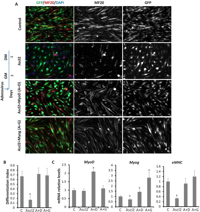
Fig. 8.
MyoD and Myog rescue Ascl2OE-induced myogenic blockage. (A) Control, Ascl2OE, Ascl2OE+MyoDOE (A+D) and Ascl2OE+MyogOE (A+G) myoblasts were induced to differentiate for 1 day. The differentiated myoblasts were stained by MF20 (red) and nuclei were counterstained with DAPI (blue). Before induction of differentiation by serum withdrawal, myoblasts were incubated with adenoviruses for 1 day, and cultured in virus-free growth medium for 1 day more. (B) Differentiation index of myoblasts, calculated by dividing the number of nuclei in myotubes (MF20+ elongated cells) by the total number of nuclei. Only GFP+ cells were used for quantification. n=3 different batches of primary myoblasts, with five different areas analyzed in each experiment. (C) Relative expression of MyoD, Myog and eMHC. Error bars represent mean with s.d. of three independent biological experiments. *P<0.05 (Student's t-test).

