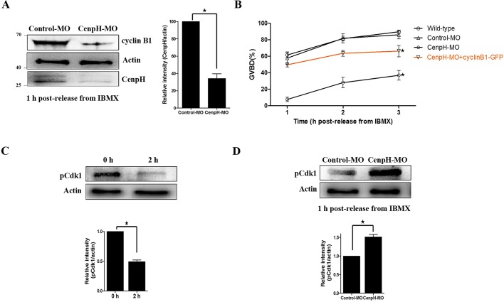
Fig. 2.
Depletion of CenpH impairs GVBD and MPF activity. (A) Western blotting of CenpH, cyclin B1 and β-actin in CenpH MO-injected and control MO-injected oocytes 1 h following release from IBMX (150 oocytes per sample). CenpH is 28 kDa, β-actin is 43 kDa and cyclin B1 is 55 kDa. The relative staining intensity of CenpH was assessed by densitometry. (B) GVBD rates at 1, 2 and 3 h following release from IBMX for wild-type, control MO-injected, CenpH MO injected, and CenpH MO+cyclin B1-GFP oocytes. (C) The phosphorylation level of Tyr15 of Cdk1 (pCdk1) in normal GV and GVBD oocytes. The relative staining intensity of pCdk1 was assessed by densitometry. (D) pCdk1 levels in control MO-injected and CenpH MO-injected oocytes 1 h following release from IBMX (150 oocytes per sample). The relative staining intensity of pCdk1 was assessed by densitometry. Data are mean±s.e.m. *P<0.05.

