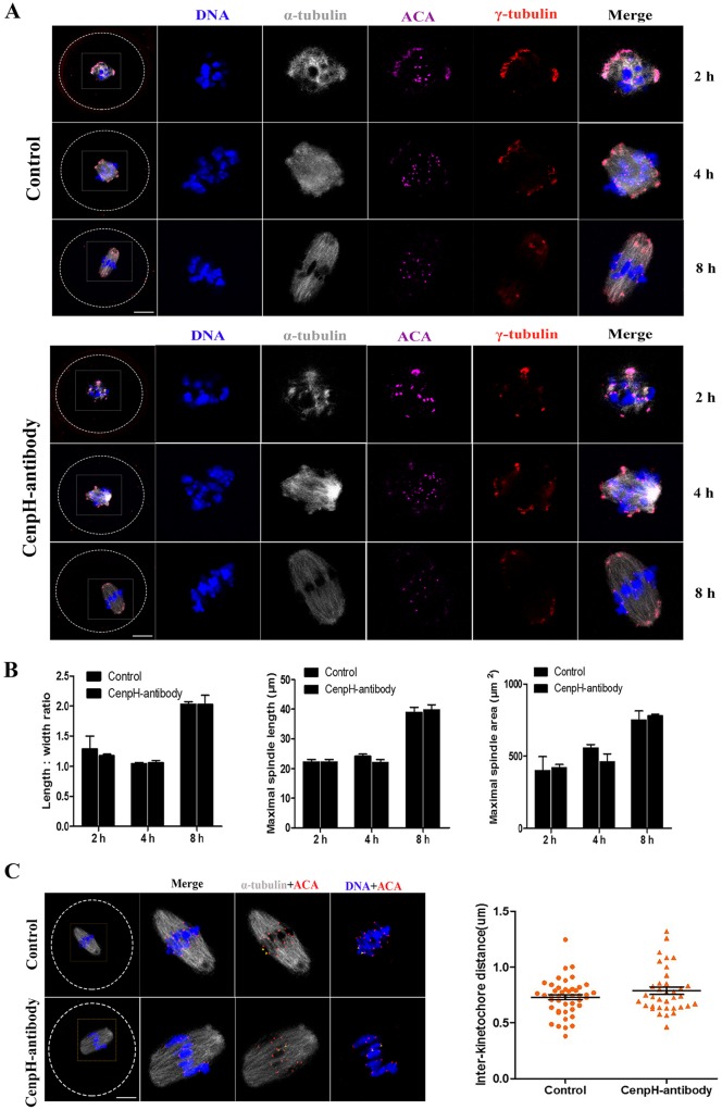
Fig. 4.
CenpH is not required for spindle assembly. CenpH antibody was microinjected into GVBD stage oocytes. (A) Confocal images of control and CenpH antibody-injected oocytes immunostained for DNA, kinetochores (ACA), microtubule organizing center (γ-tubulin) and microtubules (α-tubulin). (B) Length:width ratios of spindles, maximal spindle lengths and spindle areas for control and CenpH antibody-injected oocytes at 2 h (n=20 and n=12), 4 h (n=20 and n=14) and 8 h (n=18 and n=14). (C) Confocal images of control and CenpH antibody-injected oocytes stained for DNA and immunostained for kinetochores (ACA) and microtubules (α-tubulin) at 8 h (n=18 and n=14). The interkinetochore distance of control and CenpH antibody-injected oocytes was assessed. Data are mean± s.e.m. The total numbers of analyzed oocytes are indicated (n). Scale bars: 20 μm.

