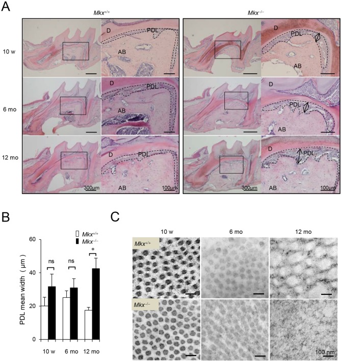
Fig. 2.
Age-related defects of the PDL in Mkx-deficient mice. (A) H&E staining of the M1 region in 10-week-, 6-month- and 12-month-old Mkx+/+ or Mkx−/− mice. D, dentin; AB, alveolar bone; PDL, periodontal ligament. Arrows indicate expansion of the PDL space. (B) Quantitative analysis for width of the PDL space in Mkx+/+ or Mkx−/− mice (n=5, 6 and 6 for 10-week-, 6-month- and 12-month-old Mkx+/+; n=3, 6, and 10 for 10-week-, 6-month- and 12-month-old Mkx−/− mice, respectively; mean±s.e.m.; ns, not significant; *P<0.05). (C) TEM analysis of collagen fibrils in the M1 furcation area. Images are representative examples.

