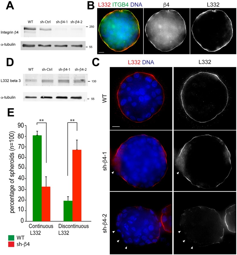
Fig. 3.
3D PIN Model – absence of α6β4 integrin expression and discontinuous laminin-332 layer. (A) Cell lysates from shRNA-expressing RWPE-1 cells were probed for β4 integrin and α-tubulin; the position of molecular mass markers is shown in kDa. (B) Day-10 WT RWPE-1 spheroids immunostained for laminin-332 (L332, red) and β4 integrin (β4, green) showing colocalization of laminin-332 and β4 integrin (yellow). (C) Day-10 RWPE-1 spheroids immunostained for laminin-332 (red) showing a continuous layer in WT RWPE-1 spheroids (top panel) and a discontinuous distribution of laminin-332 (white arrowheads) in β4 integrin-depleted spheroids (middle and bottom panels). DNA (blue). Scale bars: 10 µm. (D) Detection of the β3 chain of laminin-332 and α-tubulin in wild-type (WT), shRNA control (sh-Ctrl) and β4-integrin-depleted RWPE-1 lysates. (E) Percentage of continuous laminin-332 assembly in day-10 WT and β4 integrin-depleted spheroids. Data are mean±s.d. from three experiments, n=100. **P<0.001 (two-tailed t-test).

