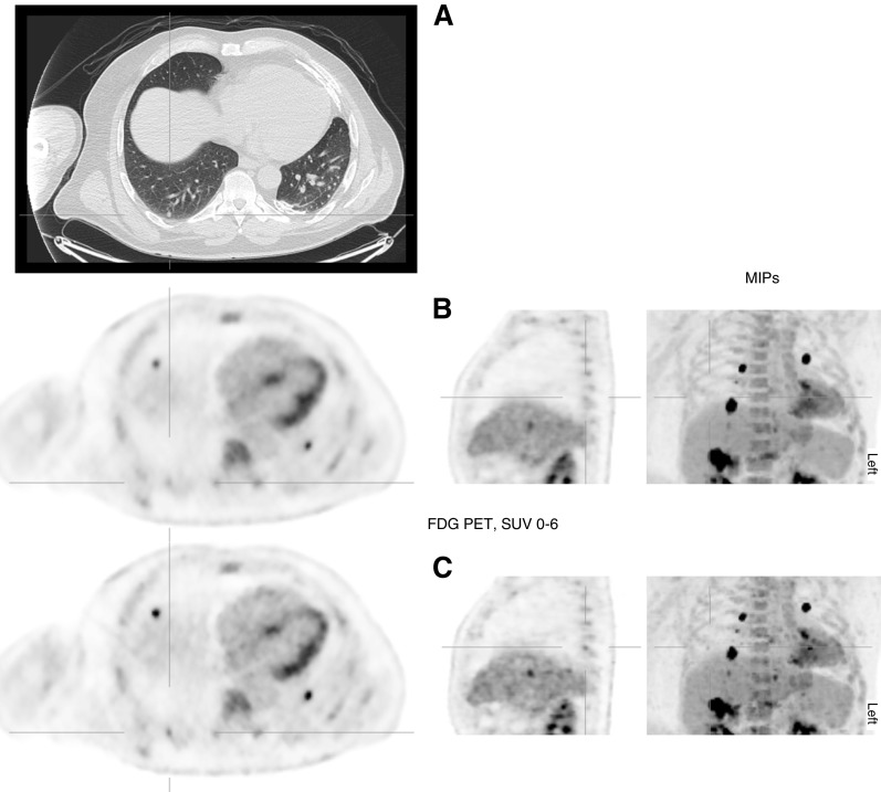
Figure 1.
Selected fludeoxyglucose F 18 (FDG) positron emission tomography (PET)–computed tomography images showing standard and data-driven gating (DDG) reconstructions. All PET images are shown on a Standardized Uptake Value (SUV) scale of 0–6. Multiple metastases are present, seen well on the maximal intensity projection (MIP), where the difference resulting from DDG is also well displayed. (A) Axial computed tomography, lung window: 6-mm pulmonary nodule posteriorly in the right lower lobe. (B) Corresponding axial, sagittal, and frontal MIPs of the PET, standard reconstruction. The nodule is not discernible from background uptake in the nearby thoracic wall. (C) Similar PET images from the DDG reconstruction. The nodule is now identifiably FDG avid, with SUVmax 2.8. Small hepatic metastases are also more conspicuous.

