Figure 3.

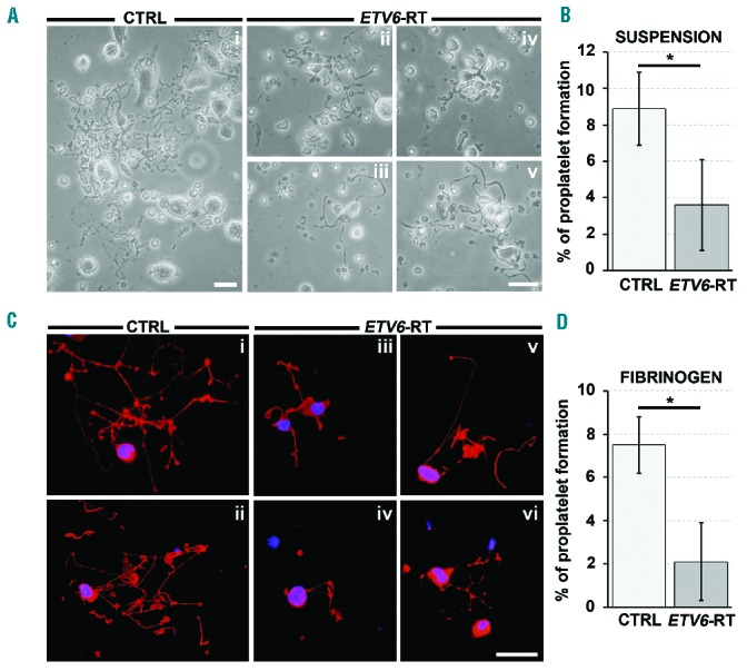
Aberrant proplatelet formation by ETV6-RT megakaryocytes. (A) Representative light microscopy analysis of proplatelet formation and structure from control (CTRL, i) and patient (ETV6-RT, ii–v) megakaryocytes cultured for 16 h in suspension (scale bar=50 μm). (B) The percentage of proplatelet-forming megakaryocytes was calculated as the number of megakaryocytes displaying at least one filamentous pseudopod with respect to total number of round megakaryocytes per analyzed field (*P<0.01). (C) Representative fluorescence microscopy analysis of proplatelet formation and structure from CTRL (i–ii) and ETV6-RT (iii–vi) megakaryocytes cultured for 16 h with adhesion on fibrinogen. The pictures clearly show defective proplatelet elongation in ETV6-RT (red=β1-tubulin; blue=nuclei; scale bar=30 μm). (D) The percentage of proplatelet-forming megakaryocytes was calculated as the number of β1-tubulin-positive cells displaying at least one pseudopod with respect to total number of round megakaryocytes per analyzed field (*P<0.01).
