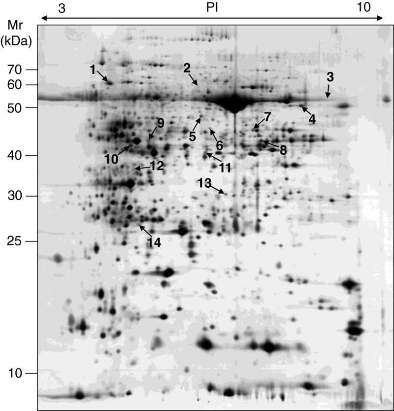
Figure 1.

A silver-stained 2-DE gel of the proteins extracted from leaf blades of both WT and spl5 mutant. For IEF, 100 μg of total proteins was loaded onto pH 3-10 IPG strips (13 cm, nonlinear), and then transferred to 12.5% SDS-polyacrylamide gel for the second-dimensional electrophoresis. The protein gel was stained with silver nitrate solution. Quantitative analysis of digitized images was carried out using the Image Master software (Amersham, USA). Arrows indicate spots with more than 2-fold change in spl5 mutant compared to WT.
