
Figure 5.
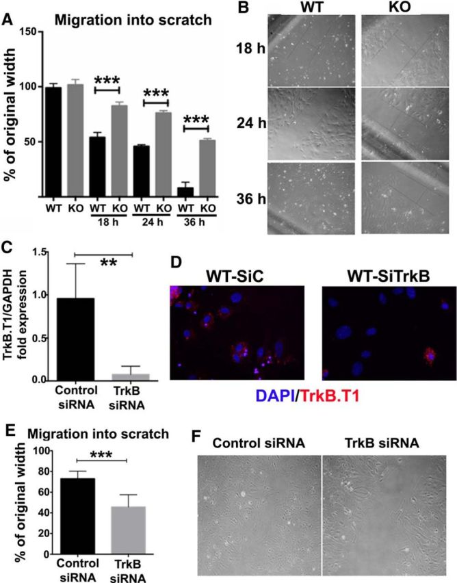
Migration into in vitro scratch wounds is impeded in astrocytes derived from trkB.T1 KO mice when compared with WT astrocytes. A, A standard scratch assay was used to examine the astrocyte's migrative capacity at 18, 24, and 36 h after injury. Compared with KO astrocytes, WT astrocytes traveled significantly farther and faster into the scratch area. N = 4 separate experiments. ***p < 0.001 versus WT [analysis with 2-tailed Mann–Whitney rank sum test (nonparametric)]. B, Representative images of WT and KO astrocyte migration into scratch areas at 18, 24, and 36 h after scratch. C–F, Acute gene deletion of trkB.T1 via transfection of astrocytes with trkB siRNA significantly reduces cellular migration into scratch wound. WT astrocytes were transfected with 10 nm of either scrambled (SiC) or targeted (SiTrkB) siRNA on 1 d postplating. Cells were harvested at day 5 postplating and mRNA isolated and cDNA prepared for real-time PCR. Real-time PCR was performed using primers against trkB.T1 (C). Immunofluorescent images showed successful transfection of cells (D). A scratch assay for migration was performed after knockdown of trkB.T1. Knockdown of trkB significantly reduced the migratory rate of astrocytes (E). Representative images of in vitro scratch assay for siRNA transfected astrocytes (F). N = 4 separate experiments. **p < 0.01, ***p < 0.001 versus Control [analysis with 2-tailed Mann–Whitney rank sum test (nonparametric)].