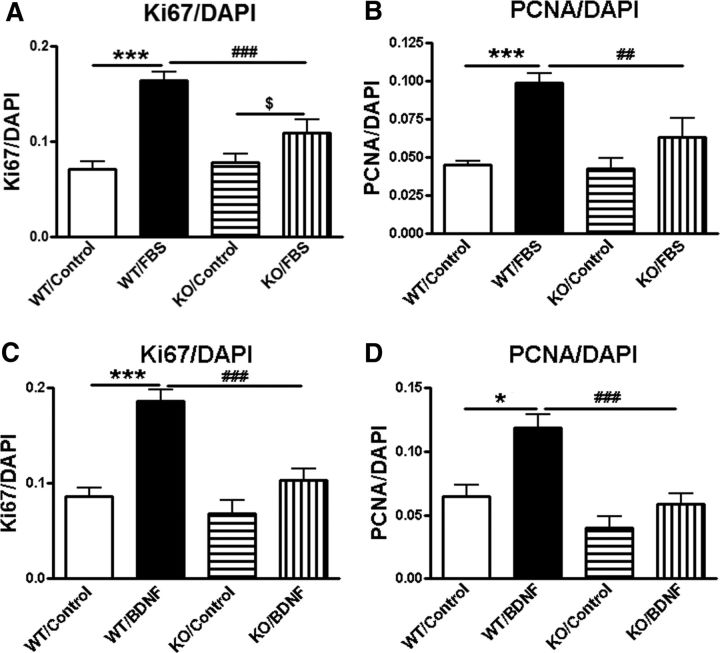
Figure 7.
Astrocytes derived from trkB.T1 KO mice demonstrate reduced proliferation in response to FBS, and are not stimulated to expand following exogenous BDNF application. A, B, Cell counts of immunolabeled astrocytes in response to 10% FBS (24 h) show slower proliferation in KO cells compared with WT cells. C, D, BDNF (50 ng/ml, 24 h) stimulates dividing of astrocytes derived from trkB.T1 WT mice, but not KO mice. N = 6 wells/group from three independent cultures. ***p < 0.001 versus WT/control; $p < 0.05 versus KO/control; ##p < 0.01, ###p < 0.001 versus WT/FBS (2-way ANOVA with Student's Newman–Keuls post hoc analysis).

