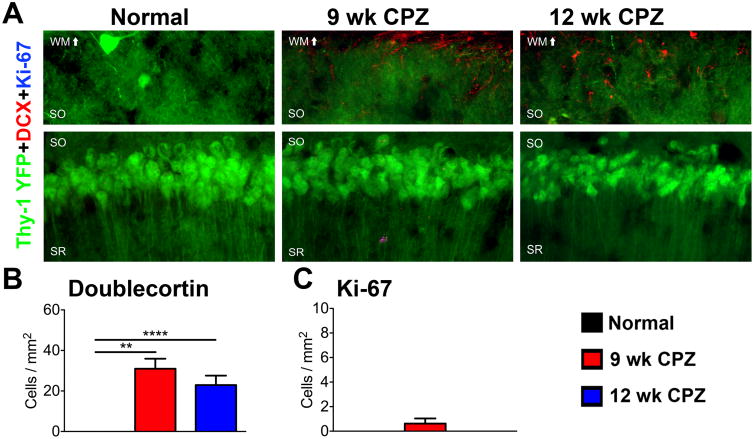
Figure 5. Status of Ki-67+ doublecortin+ cell population in the CA1 of chronically demyelinated mice with seizures.
A) Representative 40× magnified images of CA1 pyramidal cell layer immunostained for doublecortin (DCX) and Ki-67 in normal, 9 wk CPZ, and 12 wk CPZ mice. DCX+ cells with stellate morphology resembling astrocytes or microglia were observed in the CA1 SO and overlying white matter tract of 9 wk CPZ and 12 wk CPZ mice. Ki-67+ nuclei were rarely detected in the CA1 of any group, and did not co-localize with DCX. Scale bar: 10 μm.
B) DCX+ cells were significantly increased in the CA1 SO of 9 wk CPZ (****p<0.0001) and 12 wk CPZ mice mice (**p≤0.01). 8-9 animals/group, one-way ANOVA with Tukey's post test for multiple comparisons, η2=0.6077.
C) Ki-67+ nuclei counted were not significantly different between groups. 8-9 animals/group, one-way ANOVA with Tukey's posttest for multiple comparisons

