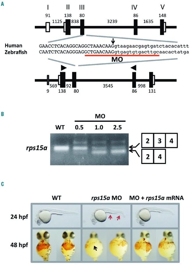
Figure 3.

Morphological features and hemoglobin staining of Rps15a-deficient zebrafish. (A) The gene structures of human RPS15A (NM_001019.4) and zebrafish rps15a (NM_212762.1). The MO target site is underlined. The arrow indicates the position of the mutated nucleotide in the patients. Arrowheads show the primer positions for the RT-PCR. Black and white boxes indicate coding and non-coding exons, respectively. Black lines indicate introns. (B) The results of RT-PCR of rps15a in wild-type and MO-injected embryos. A smaller transcript without exon 3 was observed in the morphants as seen in the patients at a comparable level. The amount of smaller transcripts correlated with the concentration of injected MOs. (C) Morphological features of wild-type and MO-injected embryos. A thin yolk sac extension and a bent tail (red arrows) were prominent in the morphants injected with 1.0 μg/μL MO targeted against rps15a, whereas these features were rescued in the embryos injected with rps15a mRNA at 400 ng/μL. Compared to the wild-type embryos, rps15a morphants injected with MO showed a drastic reduction in the number of hemoglobin-stained blood cells (black arrow). Morphants co-injected with rps15a mRNA showed recovery of the stained cells.
