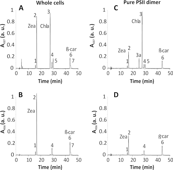
FIGURE 4.
HPLC pigment analysis of C. merolae cells and PSII dimer. Total pigments were analyzed by HPLC, and their absorbance was measured at 437 nm (for carotenoids and Chla) and at 456 nm (for carotenoids only). Peak identities are as follows. 1, Zea (cis isomer); 2, Zea; 3a, oxidized Chla; 3, Chla; 4, β-cryptoxanthin; 5, Chla′; 6, β-carotene; 7, β-carotene (cis isomer). Peaks were assigned to the corresponding pigments by LC-MS according to Ref. 39. Whole cell extract yielded a distinctive peak corresponding to Zea (A and B) nearly identical to the peak of Chla (A), whereas the β-carotene peak was 3-fold lower than that of Chla. The Zea abundance in the pure PSII dimer was significantly lower than in whole cells (C); however, the relative contribution of Zea and β-carotene was similar (D) when PSII was compared with the whole cell extract (see Table 1). a.u., arbitrary units.

