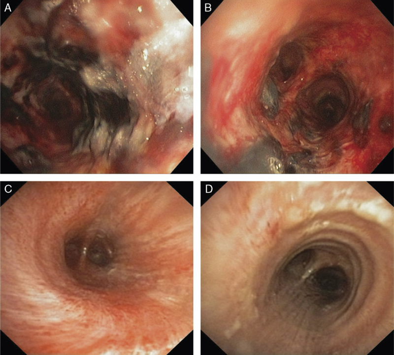
FIGURE 1.

Bronchoscopy images. On day 5, after therapy was altered to cover all pathogens, areas of mucosal hemorrhage remained in the trachea (A) and mainstem bronchus (B), but there was partial improvement seen from day 2. On day 10, a third bronchoscopy showed near-full resolution of the trachea (C) and mainstem bronchus (D).
