
Figure 2.
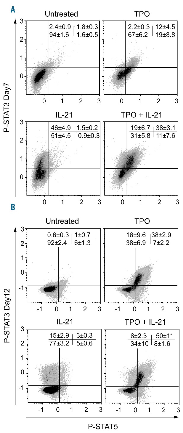
IL-21R activity changes during in vitro differentiation of megakaryocytes (MKs). CD34+ cells were cultured as described in Figure 1, scheme 1. On day 7 or 12 of culture, the cells were pre-incubated for five hours in serum-free medium without cytokines, before stimulation with thrombopoietin (TPO) and/or IL-21. The cells were then fixed, permeabilized and labeled with anti-CD41/CD61-ECD, -pSTAT3-Alexa 647 and -pSTAT5-Alexa 488 antibodies. Representative flow cytometry (FC) dot plots showing STAT3 and STAT5 phosphorylation in CD41/CD61+ cells after stimulation as indicated on days 7 and 12 of culture. Values indicate the mean±Standard Error of Mean (SEM) of the percentage of events in each quadrant in three independent experiments.