
Figure 3.
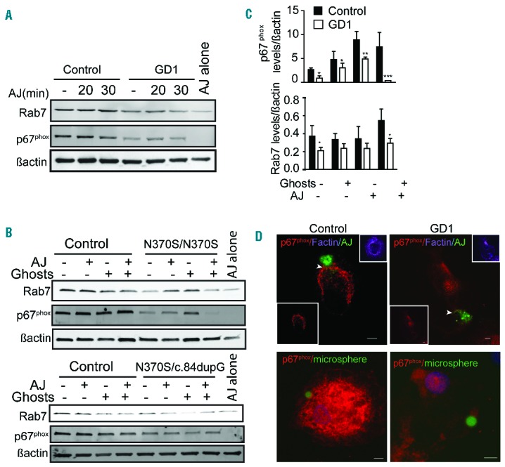
Rab7 and p67phox are reduced in Gaucher macrophages. (A, B) Control and Gaucher macrophages were co-cultured with apoptotic Jurkat cells (AJ). Macrophages were lysed after engulfing the apoptotic Jurkat cells for (A) 20, 30 min or (B) 60 min in the presence and absence of erythrocyte ghosts. Lysates were probed for Rab7 and p67phox. In (B) the upper blot shows cells from a patient with genotype N370S/N370S and the lower blot cells from a patient with genotype N370S/c.84dupG. (C) Quantification of band intensity 60 min after efferocytosis. Data represent the average ± standard deviation from three individuals with genotype N370S/N370S and two with N370S/L444P. (D) GFP-labeled apoptotic Jurkat cells (AJ) or green microbeads-PtdSer were added to control and Gaucher macrophages for 60 min, washed, fixed, and stained for p67phox (red), F-actin (purple), apoptotic Jurkat cells (AJ) or PtdSer-coated microsphere beads (green) and DAPI (blue). Z-stack images were acquired using a Zeiss 510 confocal microscope. Scale bars, 5 μm. Images represent five different independent experiments using N370S/N370S macrophages.