
Figure 5.
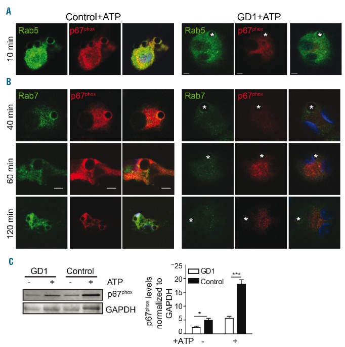
Impaired p67phox translocation to the phagosome in the presence of ATP in Gaucher macrophages. (A, B) In the presence of ATP (5 mM for 30 min), PtdSer-coated glass beads were added to control and Gaucher macrophages for different time intervals. Cells were washed, fixed and stained with p67phox (red), Rab5 or Rab7 (green) and DAPI (nuclear stain, blue), and imaged by confocal microscopy. Scale bars, 5 μm. Images represent 15 pictures at each time point from three independent experiments. (C) Cytosolic fractions from Gaucher macrophages (N370S/N370S) and control macrophages stained for p67phox in the presence and absence of ATP (5 mM for 30 min). The graph presents the results of three independent experiments from patients with genotype N370S/N370S.