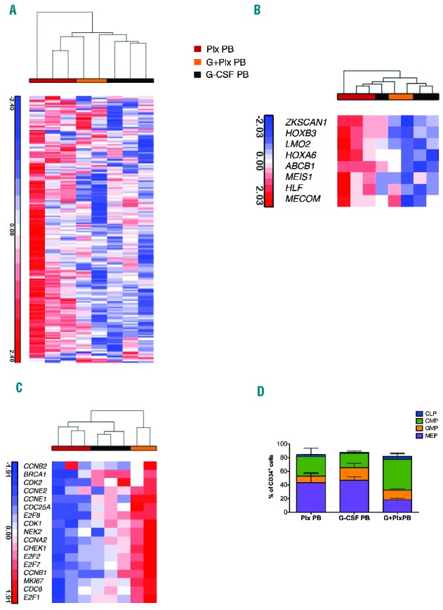
Figure 3.

‘Stemness’ signature in mobilized CD34+ cell sources. A. Hierarchical cluster analysis to assess relative distance of the transcriptome of each stem cell source. Data from mobilized stem cell sources were compared with previously generated gene sets derived from bone marrow stem and progenitor population (Online Supplemenary Figure S3). Plx PB gene expression was positively correlated with primitive gene sets. B. Heat maps of selected genes involved in HSC regulation are more highly expressed in Plx PB than in other stem cell sources. C. Heat map of regulators of cell cycle and DNA repair, showing an enrichment in the progenitor associated program in G+Plx PB expression profile. In these images, the normalized expression levels of genes are presented according to a colored gradient from the highest (red) to lowest (blue, see colored scale). D. Immunophenotypic analysis of progenitor composition in G-CSF PB, Plx PB and G+Plx PB sources. G-CSF PB (n=2); Plx PB (n=3); G+Plx PB (n=2): CMP, common myeloid progenitor (CD34+ CD38+ CD45RA− CD135+ CD10− CD7−), GMP, granulocyte-macrophage progenitor (CD34+ CD38+ CD45RA+ CD135+ CD10− CD7−), MEP, megakaryocyte-erythrocyte progenitor (CD34+ CD38+ CD45RA− CD135− CD10− CD7−), and CLP, common lymphoid progenitor (CD34+ CD38+ CD45RA+ CD10+ CD7−). The CLP, CMP, GMP and MEP subsets are reported as percentages of CD34+ cells, according to the expression of CD34, CD38, CD45RA, CD135, CD10 and CD7 surface markers. Statistically significant differences were observed by comparing the three mobilized sources with the higher CMP and the lower MEP content in G+Plx PB samples, as compared to G-CSF PB and Plx PB, respectively (P<0.05). Data are represented as mean±SEM. PB: peripheral blood.
