Figure 1.

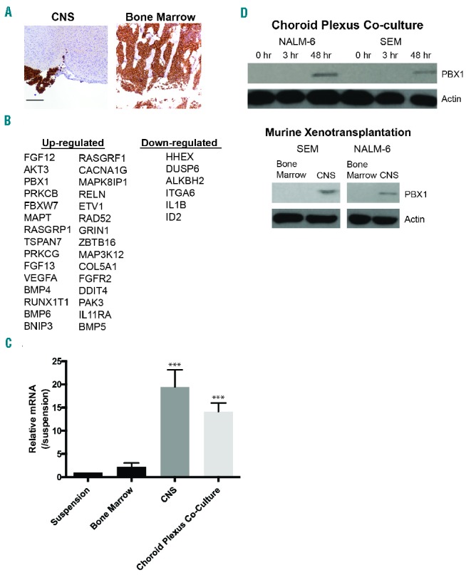
The central nervous system (CNS) and bone marrow (BM) microenvironments uniquely influence leukemia gene expression patterns. (A) Mouse brain and BM demonstrating NALM-6 leukemia involvement by human CD10 IHC staining approximately four weeks after injection with leukemia cells (10X magnification, 200 micron scale bar). Leukemia cells appear brown. (B) NALM-6 genes differentially regulated (fold change ≥2 and FDR<0.05) in the CNS niche relative to the BM identified with the Nanostring PanCancer Pathway Panel. (C) Expression of PBX1 mRNA in leukemia cells isolated from the mouse BM, CNS, or co-cultured with choroid plexus cells relative to cells in suspension determined by quantitative RT-PCR. PBX1 mRNA levels were normalized to GAPDH expression. ***P<0.0005 when leukemia cells isolated from the CNS or Z310 co-culture are compared to the other conditions. (D) Western blot showing PBX1 protein levels in NALM-6 and SEM leukemia cells grown in suspension, adherent to choroid plexus cells, or isolated from mouse CNS or BM.
