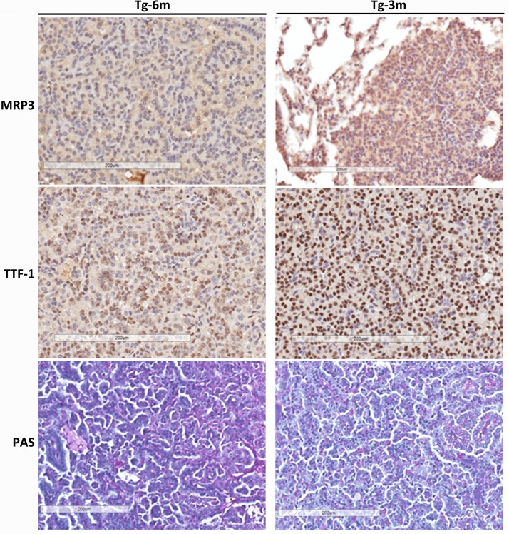
Fig 2. IHC analysis reveals lung tumors are displaying markers related to adenocarcinoma.
IHC evaluation of multidrug resistant protein 3 (MRP3), thyroid transcription factor (TTF)-1, and periodic acid-Schiff (PAS) stain in tumors obtained at 6 (Tg-6m) and 3 months (Tg-3m). MRP3 expression was only diffused in Tg-3m lung tumors. Nuclei from tumor cells of both Tg-6m and Tg-3m were stained with TTF-1; it was more intense in Tg-3m than Tg-6m tumor cells. Both Tg-6m and Tg-3m tumor samples were PAS-positive. Panels are at 200x magnification. The scale bar showed 200 μm in distance.

