
Fig. 1.
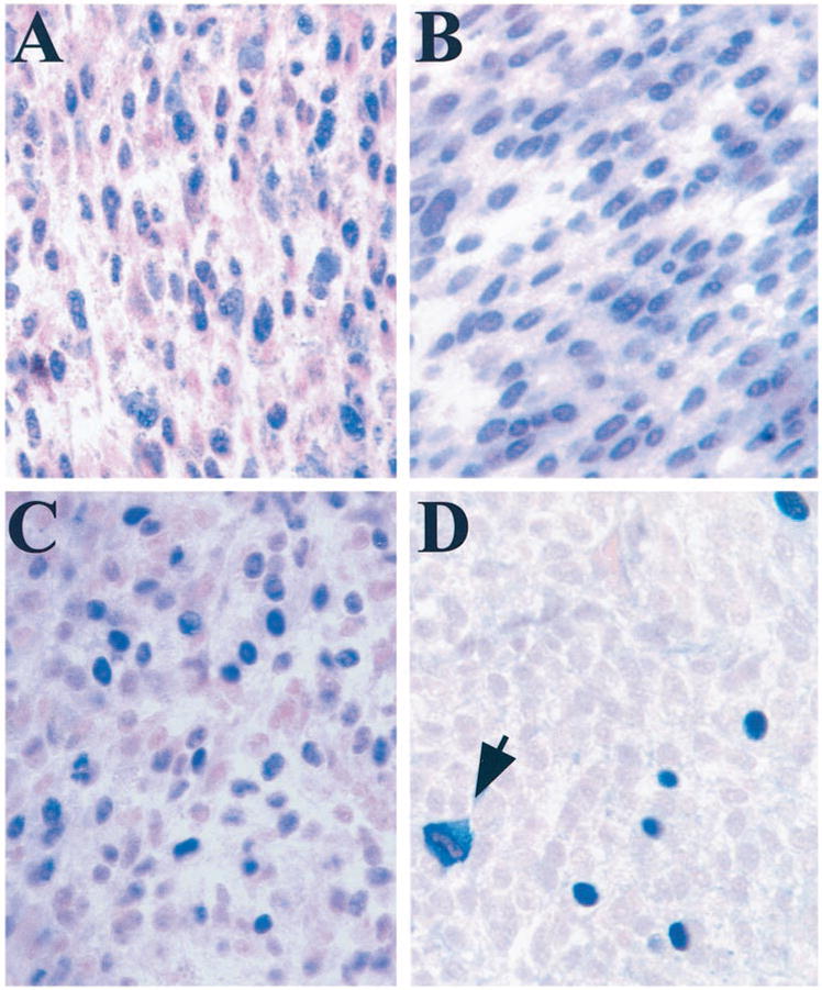
A, immunohistochemical staining of Rb in a representative uveal melanoma. Most melanoma cells had nuclear staining for Rb. ×40. B, immunohistochemical staining of p16 in a representative uveal melanoma. Most melanoma cells had nuclear staining for p16. ×40. C, immunohistochemical staining of cyclin D1 in a representative uveal melanoma. ×40. D, immunohistochemical staining for Rb phosphorylated on serine-807/811 in a representative uveal melanoma. The fraction of positive cells was similar to the fraction of cycling cells reported previously for uveal melanomas (12). Positively staining mitotic figures were observed frequently (arrow). ×40.