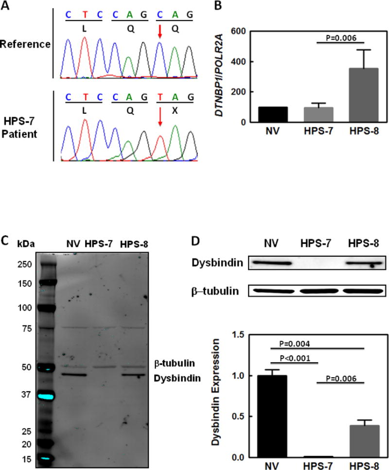
Figure 2.

Genetic and Molecular Studies in HPS-7.
(A) Sequence chromatograms showing a homozygous c.307C>T mutation (red arrow) in DTNBP1 in the proband’s genomic DNA. This nucleotide change results in the introduction of a termination codon (denoted as “X” in figure) at the protein level (Gln103*). (B) DTNBP1 mRNA expression in the proband’s fibroblasts is similar to levels in cells from a normal volunteer control (NV). Fibroblast DTNBP1 mRNA levels are increased in a patient with HPS-8 compared with control and significantly higher than in the HPS-7 patient. (C and D) Immunoblots show negligible Dysbindin protein expression in the HPS-7 proband’s fibroblast lysates compared to levels in HPS-8 or normal cells. Dysbindin protein expression in HPS-8 fibroblasts was also significantly reduced compared to normal cells. The entire immunoblot is shown in Panel C; Dysbindin and β-tubulin bands from the same blot are shown in Panel D. Loading was controlled by normalizing for β-tubulin expression in the same immunoblot; normalized Dysbindin expression in the HPS-7 and HPS-8 patients is shown relative to normal volunteer control expression.
