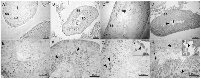
Fig 3. General view of the seminiferous tubules (A-D) and high magnification of germinal epithelium (E-H) in LEV administered groups and control.
(A-D; L: lumen; ss: spermatogenic series; sp: spermatozoa, scale bar: 50 μm). A: Normal architecture of the seminiferous tubule associated with complete spermatogenic series (ss) in control. B & C: Relatively preserved structure of seminiferous tubules and considerable number of spermatozoa in the tubular lumen in 50 mg/kg and 150 mg/kg LEV administered groups, respectively. D: Tubular degeneration with disorganisation of spermatogenic series and markedly decreased number of sperms (►) in the tubular lumen in 300 mg/kg LEV administered group. (E-H; scale bar: 20 μm). E: Intact layers of spermatogenic series in control. F: Slight vacuolation at late stage spermatids (►) in 50 mg/kg LEV administered group. G: Increased vacuolation and slight degeneration at late stage spermatids (►) in 150 mg/kg LEV administered group. H: Loss of cellular architecture in spermatogenic series (►), swelling, vacuolation (→) and necrosis (*) of the spermatids in 300 mg/kg LEV administered group. g & h: High magnification of Leydig cells (Scale bar: 10 μm). g: Vacuolation in Leydig cells (►) in 150 mg/kg LEV administered group. h: Increased vacuolation and cellular degeneration in Leydig cells (►) in 300 mg/kg LEV-administered group.

