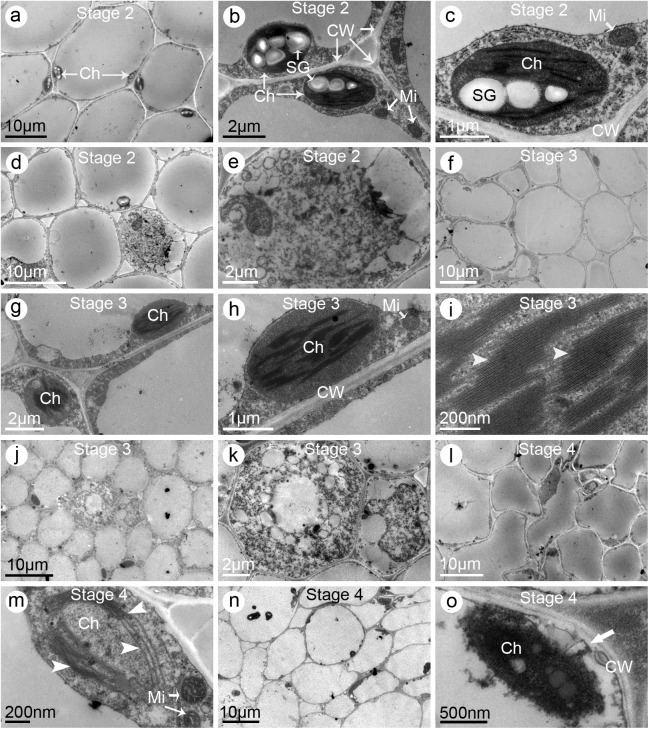
Fig 7. Electron micrographs of stigmas and styles of the single-petal (SP) jasmine plants.
(a–c) Stigmas sampled from the partially opened flowers (i.e., Stage 2). (a) Multi-cellular section exhibiting the ultrastructural characteristics. Note the starch granules in the chloroplasts. (b) Enlarged graph of (a) showing the organelles including chloroplasts, starch granules, and mitochondria. (c) A single chloroplast. (d–e) Styles sampled at Stage 2. (d) Multi-cellular section exhibiting the ultrastructural characteristics. (e) Enlarged graph of (d) showing the transfer cell. (f–i) Stigmas sampled from the fully opened flowers (i.e., Stage 3). (f) Multi-cellular section exhibiting the ultrastructural characteristics. Notice fewer starch granules were observed. (g) Enlarged graph of (f) showing several cells and their organelles. (h) Enlargement of (g) showing a single chloroplast. (i) Enlargement of (h) showing the grana and lamellae (arrow heads). (j–k) Styles sampled at Stage 3. (j) Multi-cellular section exhibiting the ultrastructural characteristics. (k) Enlarged graph of (j) showing the transfer cell. (l–m) Stigmas sampled from the flowers at one day post fully opened (i.e., Stage 4). (l) Multi-cellular section exhibiting its ultrastructural characteristics. Notice the cells were of irregular shape and intercellular spaces were larger than normal. (m) A single chloroplast showing fewer grana and lamellae (arrow heads) than normal. (n–o) Styles sampled at Stage 4. (n) Multi-cellular section exhibiting the ultrastructural characteristics. (o) A single chloroplast showing the degenerating grana and lamellae. Notice the plasmolysis (arrow) occurred. Abbreviations: Ch, chloroplast; CW, cell wall; Mi, mitochondria; SG, starch granule

