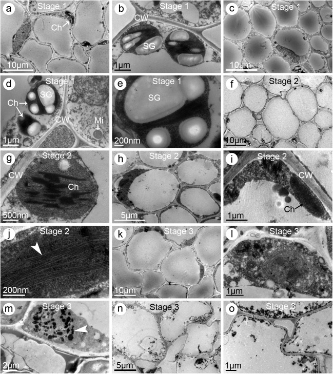
Fig 9. Electron micrographs of stigmas and styles of the multi-petal (MP) jasmine plants.
(a–b) Stigmas sampled from the flowers at one day before partially opened (i.e., Stage 1). (a) Multi-cellular section exhibiting the ultrastructural characteristics. Note many starch granules were observed. (b) Enlarged graph of (a) showing the chloroplasts and starch granules. (c–e) Styles sampled at Stage 1. (c) Multi-cellular section exhibiting the ultrastructural characteristics. Notice the cells contained dense cytoplasm. (d) Enlargement of the cell showing its organelles. (e) Enlargement of a single chloroplast with starch granules. (f–g) Stigmas sampled from the partially opened flowers (i.e., Stage 2). (f) Multi-cellular section exhibiting the ultrastructural characteristics. Note the chloroplasts were fewer than normal and starch granules were nearly disappeared. (g) Enlarged graph of a single chloroplast with irregular grana and lamellae. (h–j) Styles sampled at Stage 2. (h) Multi-cellular section exhibiting the ultrastructural characteristics. (i) Enlarged graph of (h) showing the cellular containers. (j) Enlarged graph of (h) showing the lamellae (arrow heads). (k–m) Stigmas sampled from the fully opened flowers (i.e., Stage 3). (k) Multi-cellular section exhibiting its ultrastructural characteristics. Notice no starch granules were observed in the cells. (l) Enlarged graph of (k) showing the degenerated cellular organelles. (m) Another cell showing the degraded nucleus and its apoptotic bodies (arrow heads). (n–o) Styles sampled at Stage 3. (n) Multi-cellular section exhibiting its ultrastructural characteristics. Notice the cellular organelles had entirely degenerated. (o) Enlargement of the stylar cell showing its degenerated containers. Abbreviations: Ch, chloroplast; CW, cell wall; Mi, mitochondria; SG, starch granule

