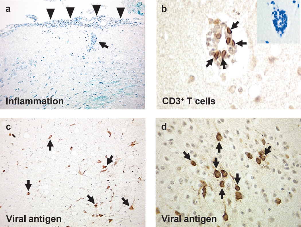
Fig. 2.
Neuropathology of West Nile virus (WNV) encephalitis. The brain was harvested from mice infected with WNV subcutaneously, and embedded in paraffin.55 (a) Meningitis (arrowheads) and perivascular cuffing (arrow) composed of mononuclear cells in the brain (Luxol fast blue stain). (b) T cells (arrows) in the perivascular cuffing by immunohistochemistry against CD3. (c, d) Viral antigens (arrows) in the cytoplasm and cell processes of neurons by immunohistochemistry with rabbit anti-WNV antibody (1 : 4,000 dilution, 81-015, BioReliance, Rockville, Maryland, USA), following trypsin treatment.

