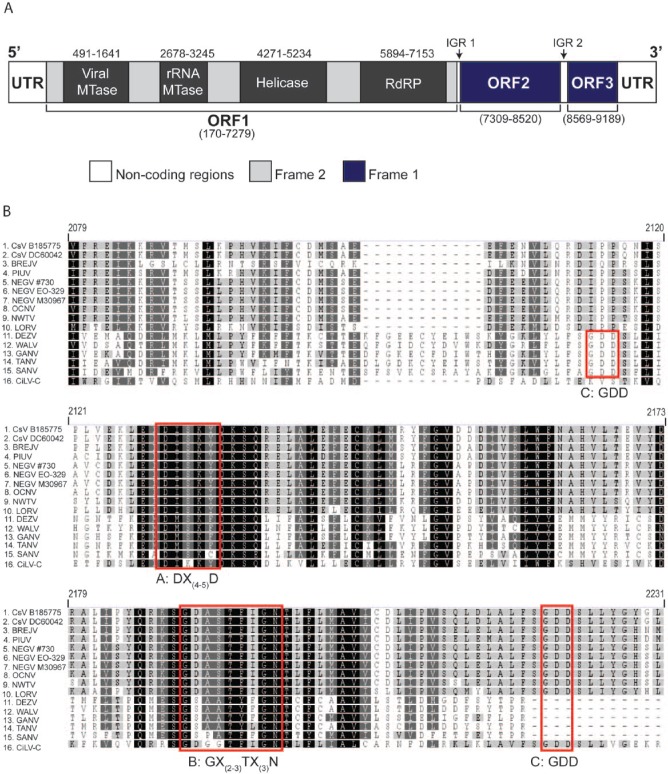
Figure 2.
Organisation of the Castlerea virus (CsV) genome. (A) A schematic of the CsV genome organisation. Regions shaded in dark blue are translated in the first frame, and light grey in the second frame and white represent regions of noncoding sequence. Intergenic regions (IGR) 1 and 2 are depicted with arrows. The black boxes represent the various replication machinery motifs within ORF1 with nucleotide positions depicted above each motif. (B) Amino acid sequence alignment of the RNA-dependent RNA polymerase (RdRP) of various negeviruses and Citrus Leprosis virus C (CiLV-C). Red boxes indicate the position of motifs – A: DX(4-5)D, B: GX(2-3)TX(3)N, and C: GDD. IGR indicates intergenic region; MTase, methyltransferase; ORF, open reading frame; UTR, untranslated region,

