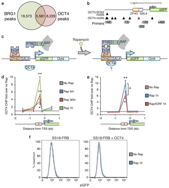
Figure 4. TOP2 is required for optimal BAF-mediated recruitment of OCT4.
(a) Venn diagram of overlapping BRG1 and OCT4 ChIP-seq peaks in ES cells. (b) Diagram of OCT4 motifs at the CiA:Oct4 locus. Red arrowhead indicates the OCT4 motif in the DNA binding 19 bp downstream from the zinc-finger recruitment site. (c) Strategy for testing BAF/TOP2 pioneering for OCT4 using BAF recruitment to the CiA:Oct4 locus in fibroblasts while expressing exogenous OCT4. OCT4 ChIP in fibroblasts with the BAF recruitment system over-expressing OCT4 treated with 3 nM rapamycin (d) in the presence of 1 μM ICRF-193 (e). (f) eGFP flow cytometry of fibroblasts over-expressing OCT4 treated with rapamycin for 1 hour. Significance assessed by t-tests as before. Lines represent means and error bars represent s.e.m. from 3 cell passages (d,e).

