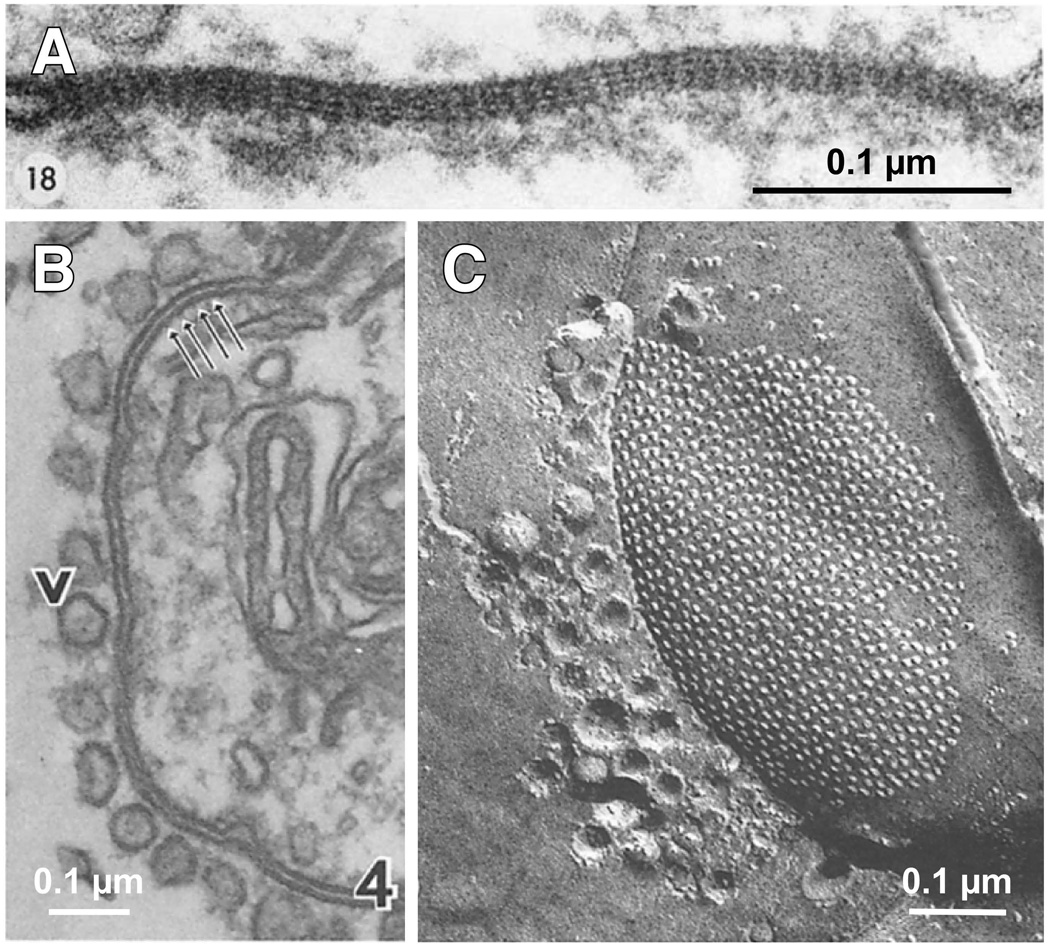
Figure 3. Structural asymmetry at electrical synapses.
Morphological asymmetries can be observed at both Cx-based and Inx-based electrical synapses. A, Electron micrograph of GJ between a Club Ending and the lateral dendrite of the Mauthner cell of a goldfish (Carassius auratus). Note the asymmetry of the ESD at each side of the junction. Modified from Brightman and Reese, 1969, with permission (Brightman and Reese, 1969). B, Electron micrograph of a GJ situated in the synaptic contact between the lateral giant axon and the giant motor fiber of a crayfish (Procambarus clarkii). The presynaptic side can be easily identified by the presence of vesicles (v), which seem to be connected to the junctional membrane. C, Freeze-fracture image (e-face) of a GJ in another crayfish electrical synapse showing the close arrangement of particles and vesicles, which are only present in the cytoplasm of the presynaptic lateral giant axon. Panels B and C modified from Hanna et al, 1978, with permission (Hanna et al., 1978). There are structural differences between vertebrate and invertebrate gap junctions including membrane separation (B) and inter-channel distance and regularity (C).

