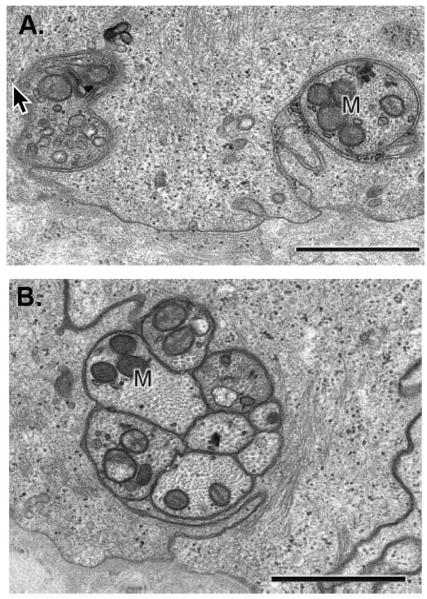
Figure 2. The SBNs contain variable numbers of individual axons bundled together.
A. Transmission electron micrograph of two cross-sectioned single nerve fibers cut in a plane perpendicular to the orientation of SBNs shown in Figure 1A. These nerves are wrapped in the basal membranes of the basal cells. B. Higher magnification of a cross-sectioned SBN bundle with 8 individual axons. This bundle is wrapped within the basolateral membranes of adjacent basal cells. Note in both A and B numerous mitochondria in the axons as indicated by the M. In addition, note that SBNs do not interact directly with the epithelial basement membrane. The cell membranes of the corneal epithelial basal cells wrap around the nerves giving the appearance in cross section that the nerves are being endocytosed by the basal cells. Images shown are taken from a paper by Muller and colleagues (2003) with permission from the publisher. Bars = 1 μm.

