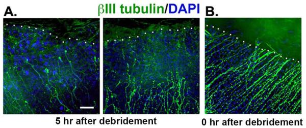
Figure 6. Proximal stubs of the SBNs dieback towards the periphery within 5 hours after injury.
Corneas wounded by 1.5 mm debridement injury and either sacrificed immediately or five hours after wounding. Tissues were stained to reveal the localization of the severed distal tips of the SBNs using an antibody against βIII tubulin (green) and the cell nuclei using DAPI (blue). A: Axons in corneas from mice sacrificed five hours after injury reveal significant retraction back from the leading edge. The white dotted line indicates the margin of the wound edge where epithelial cells are missing. The mean distance SBNs retracted was 39 μm but there is significant variation between corneas from different mice. SBNs, like myelinated PNS nerves, undergo dieback after wounding. B: Axons remain at the margin of the wound site in corneas from mice sacrificed within minutes of injury. Bar =10 μm.

