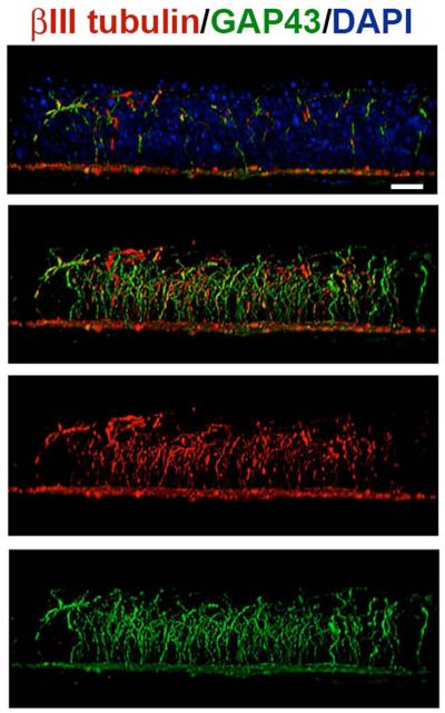
Figure 8. SBNs express the regeneration-associated protein GAP43 during homeostasis.
Unwounded corneas were stained to co-localize βIII tubulin (ρεδ) ανδ ΓΑΠ43(green); nuclei are indicated by staining with DAPI (blue) and used for whole mount imaging. Images were acquired with the 63x objective using confocal microscopy. Using Volocity, confocal image stacks are rotated to generate cross-sectional views. The field of view acquired in x and y is 135x135 μm; projection images show the full x or y field of view. The majority of the βIII tubulin+ SBNs in the unwounded cornea express GAP43 where it is localized within axons that extend apically. These data suggest that some axons are branching and growing more than others. Bar= 10 μm.

