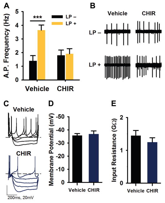
Fig. 2. GSK3 inhibition blocks light-induced increase in SCN neuronal activity.
(A) Spontaneous action potential frequencies (means ± SEM) of Per1::GFP expressing SCN neurons treated with vehicle (DMSO, 0.002%) or CHIR (1 μM) for 1 hour (ZT 23–24) following exposure to 15-min light-pulse (LP+) or no light (LP-) at ZT 22. Recordings were made 3–5 hours after onset of photic stimulus (ZT 1–3). (B) Representative cell-attached loose-patch traces (5 s) from each group in (A). ***P < 0.001, n = 41–43 cells, 3–4 slices per group. (C) Representative current clamp recordings from LP+ SCN neurons treated with vehicle or CHIR (as in A). (D–E) Means ± SEM of resting membrane potential (D) and input resistance (E) of cells represented in (C). n = 13–15 cells, 2–3 slices per group.

