
FIGURE 2.
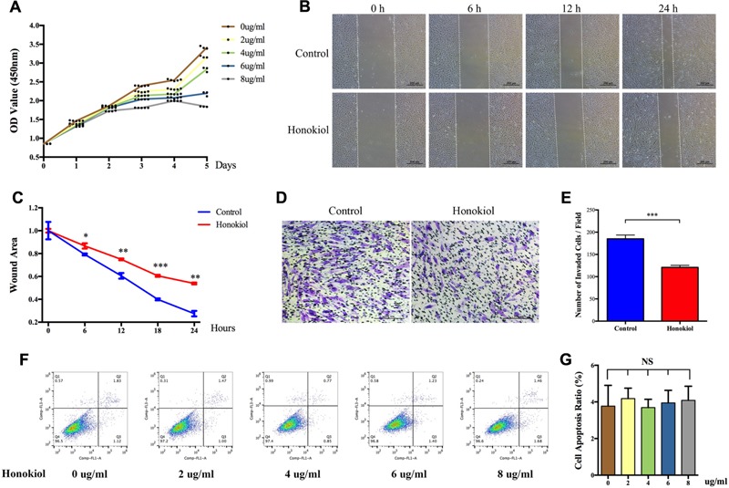
Effects of HKL on HSFs proliferation, migration, and apoptosis. (A) Proliferation curves of HSFs after treating with HKL at 0, 2, 4, 6, or 8 μg/ml for 1, 2, 3, 4, and 5 days by CCK-8 assay. (B) Representative pictures of the wound-healing assay after HKL treatment at 0 μg/ml or 6 μg/ml for 0, 6, 12, and 24 h. The white dotted lines indicate wound scratch. (C) The wound area quantified of (B). n = 3. (D) Representative pictures of the transwell assay after HKL treatment at 0 μg/ml or 6 μg/ml for 24 h. (E) The number of invaded HSFs per field quantified of (D). n = 3. (F) Flow cytometry results of cell apoptosis after treatment of HKL for 2 days at 0, 2, 4, 6, or 8 μg/ml. (G) Quantification of cell apoptosis ratio in (F). n = 3. Data are mean ± SD. ∗P < 0.05; ∗∗P < 0.01; ∗∗∗P < 0.001. OD value, optical density value; NS, no significance.