Figure 5.
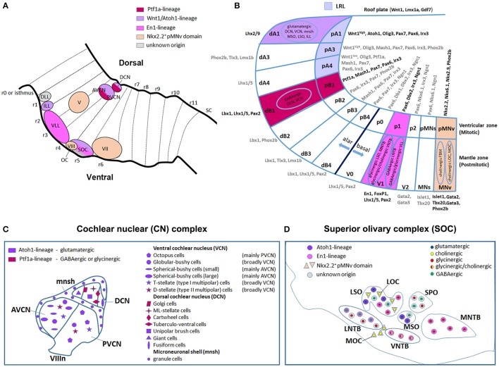
Dorso-ventral origin of auditory nuclei. (A) Schema showing a sagittal view of the hindbrain correlating the different auditory nuclei to their DV domains of origin (B) by color code. (B) Schematic transverse section through the developing hindbrain at r4–r6 levels, showing the DV domain of progenitors and derived neurons and their contribution to auditory nuclei, modified from Nothwang (2016). Note that the dA3 domain is absent in r1–r3, while dA4, dB2, dB3, and dB4 are missing in r1 (Sieber et al., 2007; Gray, 2008). The pMNs domain, dorsal to the pMNv, is only present in r1 and r5 (Takahashi and Osumi, 2002; Guthrie, 2007). (C,D) Schematic representation of the DV origin and neurotransmitter phenotype of auditory neuronal populations: (C) cochlear nuclear complex [AVCN, PVCN, DCN and microneuronal shell (mnsh)], (D) superior olivary complex (LSO, MSO, MNTB, VNTB, LNTB, SPO) and olivocochlear (LOC and MOC) neurons, modified from Fujiyama et al. (2009) and Altieri et al. (2015), respectively. The same color code is maintained from (A–D).

