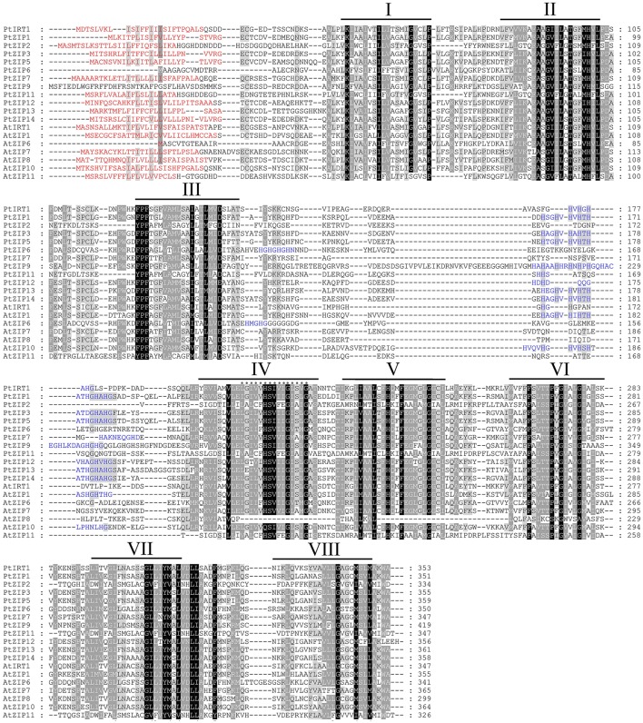
Figure 1.
Multiple sequence alignment of PtZIP and AtZIP proteins. Twelve PtZIP proteins isolated from trifoliate orange and seven representative AtZIP proteins from Arabidopsis were aligned using ClustalW. Similar amino acids are indicated by dark or light shading, while signal peptides in the N-terminal end are highlighted in red. Transmembrane (TM) domains were shown as lines above the sequences and numbered I to VIII. The variable region rich in histidine residues between TM-III and TM-IV is highlighted in blue. The 15 ZIP signature sequences in TM-IV domain are marked with asterisks.

