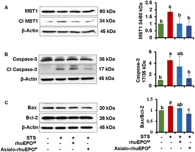
FIGURE 4.

Western blot of MST1 (A), Caspase-3 (B), Bax and Bcl-2 (C). The levels of these proteins were measured in cell lysates prepared from cells treated with PBS containing 0.1% BSA (vehicle control), 0.123 μM STS, 0.123 μM STS+60 IU/ml rhuEPOM or 0.123 μM STS+60 IU/ml asialo-rhuEPOP. Active MST1 and caspase-3 were detected using an anti-MST1 and anti-caspase-3 antibody, respectively, which also cross-react with proMST1 and procaspase-3. Bax and Bcl-2 specific antibodies were used to detect these proteins. β-Actin was used as internal control. The experiment was repeated twice. All data plotted are the average of two independent experiments ± SD. Different letters labeled represent significant difference at p < 0.05 level.
