Figure 1.
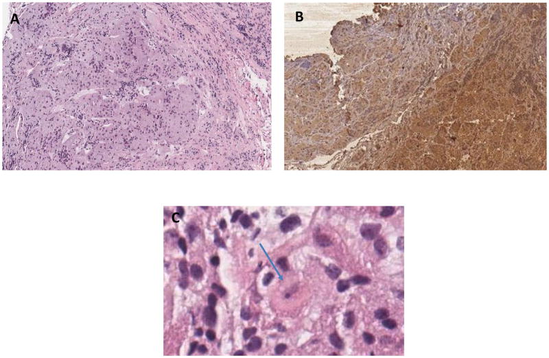
Histological examination of the pituitary gland demonstrates areas of architectural distortion and acinar expansion with displaced reticulin network, staining positively for ACTH, with adjacent normal pituitary tissue. On Panel C, a few cells showed Crooke’s changes with replacement of cytoplasmic granules of basophil cells by homogeneous hyaline material (see arrow).
A. Pituitary Adenoma hematoxylin and eosin (H&E) 4×
B. Pituitary Adenoma with Immunostaining for ACTH 4×
C. Pituitary Adenoma H&E 40 x,
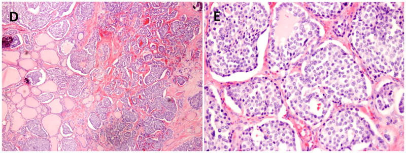
Histological examination of the thyroid gland demonstrates medullary thyroid carcinoma with characteristic nests of spindle shaped cells and amyloid deposits.
D. Medullary carcinoma of the thyroid 10×
E. Medullary carcinoma of the thyroid 20×


