Figure 8.
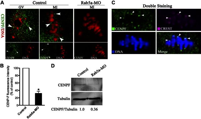
Rab5a Depletion reduces the CENPF localization/levels on kinetochores in oocytes. A) Control and Rab5a-MO oocytes were labeled with anti-CENPF antibody (green) and counterstained for chromosomes (red). Representative images of GV and MI oocytes are shown; arrowheads indicate CENPF localization. B) Quantification of CENPF staining in control and Rab5a-MO oocytes; 30 oocytes/group were analyzed. C) Double staining of metaphase oocytes with CREST antibody (red) and CENPF antibody (green), and counterstaining of chromosome with Hoechst 33342 (blue), confirming the CENPF puncta colocalization with kinetochores (arrowheads). D) Western blot showing the dramatically reduced CENPF protein levels in oocytes following Rab5a knockdown, with tubulin as a loading control. Bars represent means ± sd. *P < 0.05 vs. controls.

