
Fig. 3.
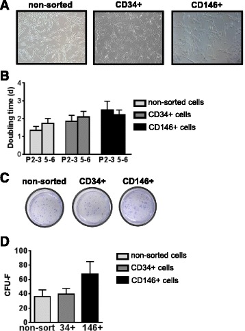
Growth of sorted cells in culture. a Micrographs showing similar morphology for non-sorted, CD34+, and CD146+ cells grown in DMEM supplemented with 20% FBS and passaged at a density of 5000 cells/cm2. b Doubling times (days) for non-sorted, CD34+, and CD146+ cells at passages 2–3 (P2-3) and 5–6 (P5-6). n ≥ 5; mean ± SEM. c, d Colonies obtained from cells grown at low density (5 cells/cm2) were stained with crystal violet (c) and CFU-F were counted (d). n ≥ 3; mean ± SEM. CFU-F colony-forming unit fibroblasts