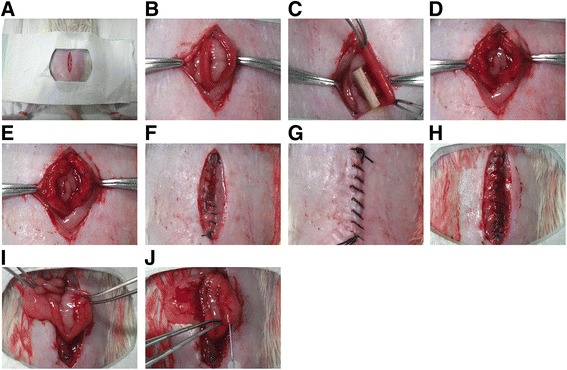
Fig. 1.

Establishment and treatments of a rat model of uterine scars. a-b After all rats were anesthetized by intraperitoneal injection of diazepam (50 mg/kg) and ketamine (50 mg/kg), a low abdominal midline incision was made to expose uterine horns. c-d The uterine wall of approximately 1.0 cm in length and 0.5 cm in width (one-third of the uterine circumference) was excised from each uterine horn, and the mesometrium was retained. e The four margins of the uterine wound were marked using a 6-0 nylon suture. f-g After rinsing the abdominal cavity with saline, the rectus fascia and skin were sutured with a 4-0 silk suture in an interrupted fashion. h–i Thirty days after full-thickness excision of uterine walls, a second incision was made in the abdominal wall of each rat to confirm scar formation. j PBS, degradable collagen fibres, UC-MSCs or UC-MSCs mixed with gelatinous degradable collagen fibres were injected into four pre-marked points surrounding each uterine scar
