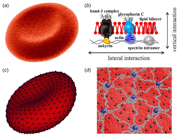
Fig. 1.

(a and b) Schematic representation of a healthy human RBC (a) and its complex membrane structure (b). The cell membrane is made of a lipid bilayer reinforced on its inner face by a flexible two-dimensional spectrin network. (c and d) Schematic view of the particle-based whole-cell model (c) and composite membrane model (d). For the whole-cell model (c), the lipid bilayer and cytoskeleton are rendered in dark gray and black triangular networks. For the coarse-grained composite membrane model (d), the dark gray, black, and light gray particles represent clusters of lipid molecules, actin junctions, and spectrin filaments of cytoskeleton, respectively; the black particles signify band-3 complexes.
