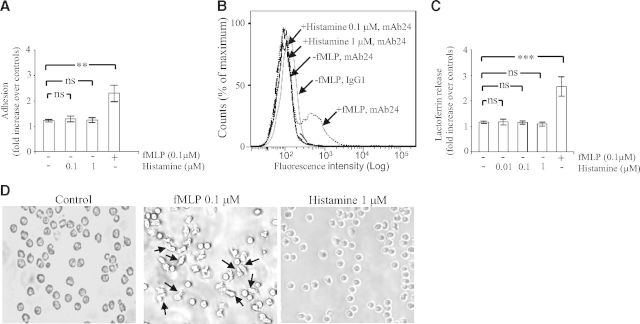
Figure 1. Histamine does not induce a Mac-1-dependent adhesion and degranulation.
(A) Human PMNs (0.5×10⁶) were incubated on plates coated with fibrinogen in the absence (−) or presence (+) of fMLP (0.1 μM) or histamine (0.1–1 μM). After 30 min, nonadherent cells were removed by aspiration, and adherent cells were fixed with paraformaldehyde and stained with crystal violet. Subsequently, the OD of the eluted dye was read by spectrophotometry at 570 nm. Adhesion is expressed as fold increase over nonstimulated control cells incubated for 30 min on BSA-coated plates (taken as 1 U). The data represent means ± sem of four separate experiments. Measurements for each experimental condition were performed in triplicate. (B) Human PMNs in suspension, stimulated or not with fMLP (0.1 μM) or histamine (0.1–1 μM), were incubated for 30 min at 37°C in the presence of the mAb24 (1 μg/ml) or an isotype-matched control IgG1 antibody (1 μg/ml). Thereafter, cells were collected, washed, and resuspended in medium containing FITC-labeled anti-mouse IgGs. Representative flow cytometry histograms (out of at least three), depicting mean fluorescence intensity of mAb24 epitope expression on human PMNs, are shown. (C) Human PMNs (1×10⁶) were incubated on plates coated with fibrinogen in the absence (−) or presence (+) of fMLP (0.1 μM) or histamine (0.01–1 μM). After 30 min, the concentration of lactoferrin in the extracellular milieu was measured by ELISA as described in Materials and Methods. The data represent means ± sem of six separate experiments. Lactoferrin release is expressed as fold increase over nonstimulated control cells incubated for 30 min on BSA-coated plates (taken as 1 U). Measurements for each experimental condition were performed in triplicate. (D) PMNs were incubated on plates coated with fibrinogen in the absence (control; left) or presence of fMLP (0.1 μM; middle) or histamine (1 μM; right). After 30 min, adhered cells were visualized using a Leitz Diaplan microscope and a 5-megapixel Leica DFC420 digital camera (Leica Microsystems, Buffalo Grove, IL, USA). Arrows show polarized and elongated PMNs. **P < 0.01, and ***P < 0.001; ns, not significant by one-way ANOVA with post hoc Bonferroni's test compared with controls.

