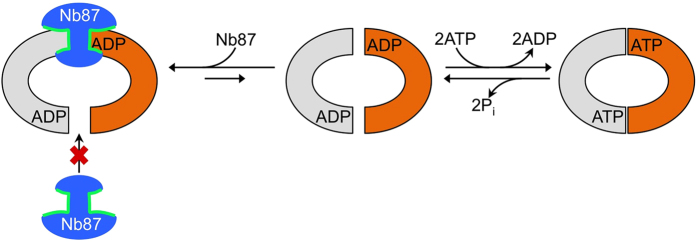
Figure 4. Schematic showing a cytoplasmic view of the “sticky-doorstop” inhibitory mechanism of Nb87.
Orange and grey shapes depict the NBDs of PglK. The middle and right panels interconvert during productive ATPase and flippase cycles. PglK subunits are represented in colors grey and orange; Inhibitory Nb87 is represented in blue and its CDR loops in green.

