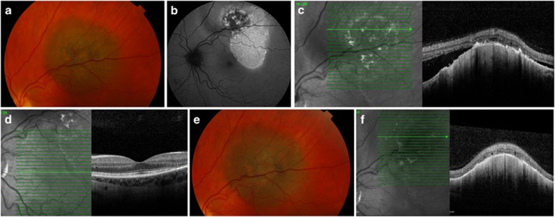
Figure 2.
Pigmented choroidal lesion (a; patient number 8 in Figure 1), 2.5 mm from the optic disc, with scattered lipofuscin orange pigment, corresponding to areas of hyper-autofluorescence (b). Optical coherence tomography demonstrated SRF over the lesion (c), but not over the fovea (d). Sixteen months after first PDT session, the lesion is stable in size (e) and SRF eliminated (f).

