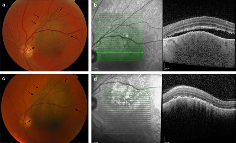
Figure 3.
Pigmented choroidal melanoma (a; patient number 3 in Figure 1), 0.5 mm from the optic disc, with scattered orange pigment and overlying SRF (b). The patient was treated with three PDT sessions; however showed tumor radial enlargement (c), detected 5 months after first and 3 months after the last PDT session. Note that despite treatment failure SRF over the lesion was eliminated (d). The patient was thereafter successfully treated with a notched plaque.

