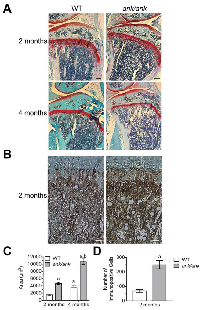
Figure 1.
A: Histological analysis of the bone marrow of 2-month and 4-month old WT and ank/ank mice (tibia, safranin O staining). Note the increased fat fraction in the bone marrow of 2-month and 4-month old ank/ank mice compared to WT littermates. Bar, 200 μm. B: Immunohistological staining with antibodies specific for perilipin, a protein that coats lipid droplets in adipocytes, of sections from the bone marrow of 2 month-old WT and ank/ank mice. Bar, 200 μm. C: Quantitative analysis of the total area (μm2) of the fat vacuoles in the bone marrow of 2-month and 4-month old ank/ank and wild type tibiae D: Number of cells immunopositive for perilipin in an area 100μm to 700μm distal to the growth plate. Sections from three different ank/ank mice and three different wild type littermates were analyzed. Data are expressed as mean ± SD. (ap < 0.01 vs. 2-month old WT tibia; bp < 0.01 vs. 2-month old ank/ank tibia).

