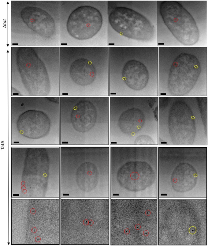
Figure 2. Electron micrographs of E. coli cells, overexpressing TatA, immunogold-labelled following primary antibody detection against TatA protein.
Ultrathin sections of E. coli cells overexpressing TatA were immunolabelled using a polyclonal antibody raised against TatA (shown in rows 2–4, with row 5 showing close-ups of individual gold particles from row 4). TatA was found to exhibit a random distribution in inner membrane (yellow circles) and was also present in the cytoplasm (red circles). Controls: cells that did not express Tat machinery were immunolabelled in the same manner. These cells largely lacked any gold binding, although a few cells bound gold in the cytoplasm and very sparingly at the inner membrane (representative images [labelled (Δtat)] in the bottom panel). Images were taken on JEOL-2010F TEM at a 12 000× magnification. Scale bar = 200 nm.

