
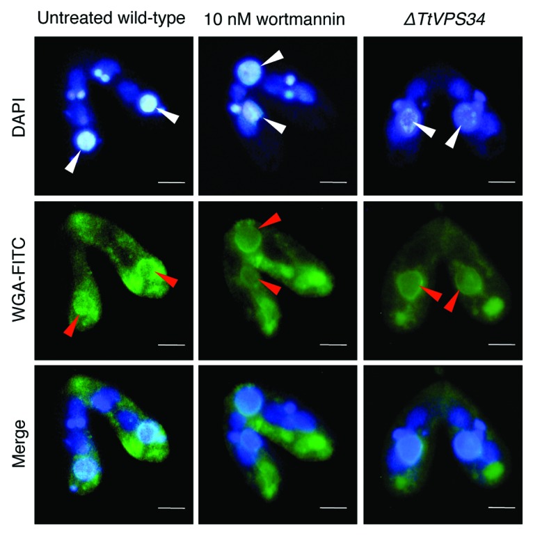
Figure 6. Binding of lectin to the parental macronucleus. Conjugating cells at 8 h were fixed and stained with DAPI (upper) and FITC-labeled WGA (middle). The lower parts show a merged image. White arrowheads, parental macronucleus; red arrowheads, concentrated FITC-signal on the nuclear surface. Scale bars: 10 μm.